Surface relaxivity & the myelin water fraction
The claim, in one line: every microstructure fit silently assumes water relaxes at a single bulk \(T_2\) — but water relaxes faster where it touches a wall, and intra- and extra-axonal water touch different amounts of wall. That \(S/V\)-dependent shortening biases the intra-axonal signal fraction and the myelin water fraction before any estimator sees the signal.
This page shows the effect the way the accompanying preprint does — "Surface relaxivity biases diffusion and relaxometry microstructure estimates" (source + PDF, reproducible with the public engines) — and lets you rerun it.
The physics
Water at a permeable boundary loses transverse magnetisation on contact (Brownstein–Tarr). The apparent rate a multi-echo fit sees is a bulk rate plus a surface rate,
with \(\rho \approx 1.16\,\mu\)m/s the cited surface relaxivity (Barakovic et al. 2023). Because \(\rho\,(S/V)\) is b-independent and isotropic, it is inseparable from bulk \(T_2\) — you cannot fit it away, and it rides through the \(b{=}0\) every fraction estimator normalises by.
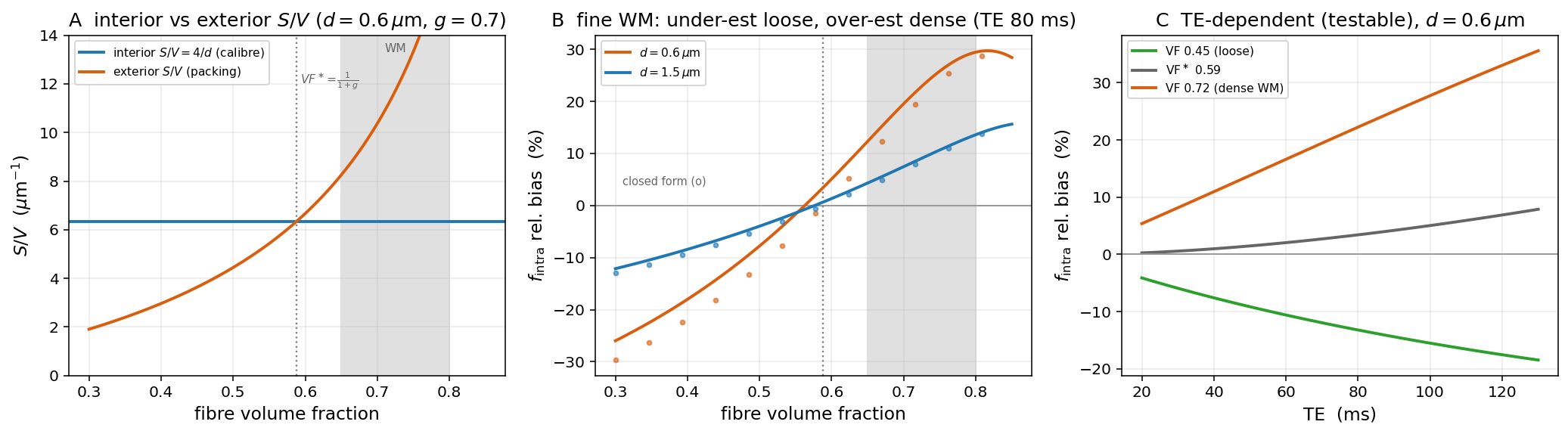
The two water pools have different geometry, hence different \(S/V\): a small, densely packed axon presents more extra-axonal wall per unit volume than a big one. The interior/exterior ratio is
independent of the calibre distribution. Physiological white matter (fibre volume fraction \(\sim0.65\)–\(0.80\)) sits above this crossover, so the exterior wins and surface relaxivity over-weights the intra-axonal signal.
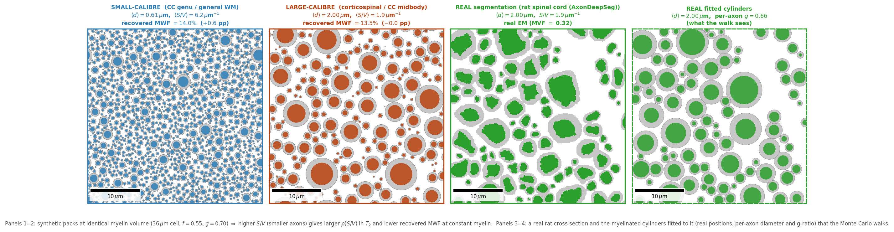
See it happen (dmipy-sim)
A CPMG train on the canonical white-matter substrate, one Monte-Carlo walk, every walker coloured by its magnitude \(|M|\). Without surface relaxivity each pool would slide down as a single spike at its bulk-\(T_2\) rate; surface relaxivity gives every walker its own wall-contact history and pushes it below that ceiling — intra-axonal (higher \(S/V\)) falls ~4 %, extra-axonal only ~1 %. That gap is the apparent-\(T_2\) shortening the fit misreads.
More of the pedagogy (phase, spatial view) is on the Pedagogy page.
What it does to your numbers
Diffusion — the intra-axonal fraction. Above the packing crossover, surface relaxivity over-weights the intra-axonal signal by \(\approx 12\%\) over the robust packing band at clinical PGSE (\(\mathrm{TE}=80\) ms, cited \(\rho\)). It under-estimates in loosely packed tissue and over-estimates in dense white matter, crossing zero at \(\mathrm{VF}^\ast=1/(1+g)\) — a closed-form sign law with a testable, packing-dependent TE drift.
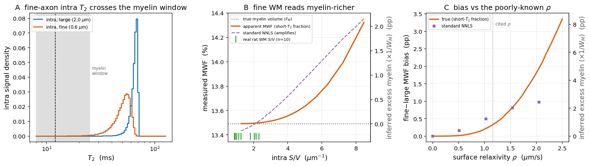
Relaxometry — the myelin water fraction. The same physics reads through a T₂ spectrum as a smaller MWF bias: the thinnest axons' intra-water crosses below the ~25 ms myelin window and is counted as myelin, so fine white matter reads myelin-richer (~0.33 pp at cited \(\rho\)). That is far beneath single-voxel noise and only exposed by regional averaging — but it is super-linear in the order-of-magnitude-uncertain \(\rho\).
Why this matters even though it's small
The MWF bias is sub-noise per voxel, but it is a spatially structured systematic — it tracks packing, so it does not average away across a region or a cohort, and it scales with a relaxivity known only to an order of magnitude. The \(f_{\mathrm{intra}}\) bias is first-order and packing-dependent. Neither shows up unless you put the surface term in the model.
Reproduce it from the public pair
Simulate a CPMG echo train (one walk) with dmipy-sim, then fit the myelin-water fraction with dmipy-fit — per-compartment \(T_2\) baked into the geometry, no private code:
import numpy as np
import dmipy_sim as ds
from dmipy_fit.white_matter.mwf import t2_spectrum_mwf

geom = ds.MyelinatedCylinder(inner_radius=2.5e-6, outer_radius=3.57e-6, orientation=(0, 0, 1),
    D_intra=1.7e-9, D_extra=1.7e-9, D_myelin=0.1e-9,
    T2_intra=0.080, T2_myelin=0.015, T2_extra=0.080)

echo_times = np.arange(1, 33) * 8e-3
S = np.array([
    float(np.asarray(ds.simulate(
        40_000,
        waveform=ds.pgse(delta=te / 2 - 1e-4, DELTA=te / 2, G_magnitude=0.0,
                         bvecs=[[0, 0, 1]], n_t=400),
        geometry=geom, seed=1)).ravel()[0])
    for te in echo_times])
mwf, T2_grid, spectrum = t2_spectrum_mwf(S / S[0], echo_times)   # NNLS T2 spectrum -> MWF
Both engines take the outer (fibre) diameter Gamma distribution; the analytical model derives the exterior \(S/V\) from it with the same closed form the simulator walks, so a fit and a simulation built from the same parameters describe the same tissue:
from dmipy_fit.white_matter.composition import build_white_matter_model
model, params = build_white_matter_model(gamma_shape=2.0,
                                         gamma_scale_outer_diameter=0.30e-6,
                                         f_axon=0.55, rho2=15e-6)
The correction the paper proposes is a single-TE, closed-form odds rescaling that injects a histology \(S/V\) prior — it harmonises fractions across cohorts scanned at different echo times. See the preprint for the full derivation, the demyelination-trajectory result, and the Monte-Carlo validation.